Friday, June 13, 2008

Google Health for Medical Records

Google views its expansion into health records management as logical because its search engine already processes millions of requests from people trying to find information about injuries, illnesses and treatments. Before this public launch, Google stored medical records for a few thousand patients at the nonprofit Cleveland Clinic.

Google's online filing cabinet for medical records opened to the public, giving users instant electronic access to their health histories and worrying a privacy advocate.

Called Google Health, the service lets users link information from a handful of pharmacies and care providers, including Quest Diagnostics labs. Google plans to add more.

Similar offerings include Microsoft Relevant Products/Services Corp.'s HealthVault and Revolution Health, which is backed by AOL co-founder Steve Case.

Google Health differentiates itself from the pack through its user interface and things like the public availability of its application program interface, or API, said Marissa Mayer, the Google executive overseeing the service.

Mary Adams, 45, a Cleveland Clinic patient who participated in the Google Health pilot, said that she was initially concerned about the privacy of her medical information.

Still, she felt safe enough to enroll and has been using the service for about six months, linking it with an online health management tool from the Cleveland Clinic and adding information on prescriptions and doctors to her online profile.

"I hate pieces of paper lying around my house, so I love the fact that i can log on with my normal Google login info and see everything at a glance," she said, adding that with its public availability she'll try to get her sister to use it.

The service, still a non-final "beta" version, does not include ads. But Mayer said Google doesn't plan to start placing them to support the site. A search box on Google Health pages leads to standard Google search results pages, where there are advertisements.

Besides importing records from providers, users can enhance their password-protected profiles with details such as allergies and medications, they can search for doctors and they can locate Web-based health-related tools.

Mountain View-based Google Inc. views its expansion into health records management as logical because its search engine already processes millions of requests from people trying to find information about injuries, illnesses and recommended treatments.

Before this public launch, Google stored medical records for a few thousand patient volunteers at the nonprofit Cleveland Clinic.

The health venture provides fodder for privacy watchdogs who believe Google already has too much about the interests and habits of its users in its logs of search requests and its vaults of e-mail archives.

Pam Dixon, executive director of the World Privacy Forum, said services like Google Health are troublesome because they aren't covered by the Health Insurance Portability and Accountability Act, or HIPAA.

Dixon's group issued a cautionary report on the topic in February on such third-party services.

Passed in 1996, HIPAA set strict standards for the security of medical records. Among other things, the law requires anyone seeking a patient's records by subpoena to notify the patient and give the patient an opportunity to fight the request.

By transferring records to an external service, patients could unwittingly make it easier for the government, a legal adversary or a marketing concern to obtain private information, Dixon said.

"We are in uncharted territory here. A privacy policy, I don't think, is enough to protect what needs to be protected in a doctor-patient record," Dixon said.

Mayer said, however, that users medical records "are generally speaking as safe with Google as they would be with a HIPAA-regulated entity."

During a webcast Monday, she said users' health information is stored at Google's "highest level of security" on computers that are more secure than those used for the company's search functions.

Mayer said in an interview with The Associated Press that Google will not aggregate users' health information across services so activity on the health service will not show up in search results.

Monday, June 2, 2008

Blocked Nose In Infants

Blocked Nose In Infants

Blocking of nose is common in babies under six months old. It is usually due to normal mucus that collects in the nose, which is difficult for the baby to clear.

Causes

Blocking of nose is not caused by colds or infections. However an infection can make things from bad to worse. Feeding can become difficult if the baby cannot breathe well due to blocked nose.

General measures


Nothing needs to be done if the baby is comfortable and able to feed. If feeding becomes difficult, the following may help.

Gently stroke the inside of a nostril with a wisp of cotton wool. This may make the baby sneeze and clear the blocked mucus. This method may help clear the mucus from the baby's nostrils before feeds and before sleep.

Increase the humidity of the room where the baby sleeps by placing a bucket of warm water. This raises the humidity, which may help to loosen thick mucus.

Saline drops (salt water drops) may be useful if the above measures do not help. Saline drops thin out the mucus and make it easier for the baby to clear from the nose. Use saline drops just before feeds when the nose is blocked. If saline is used too often, the skin around the nose may become a little sore.

Ephedrine drops prescribed by your doctor is very good at drying up secretions in the nose. But ephedrine drops should only be used 2-3 times a day for a few days only. Rebound nasal congestion may develop if ephedrine drops are used frequently every day for more than a week, which can make the blocked nose worse.

Tests for Glucose

Tests for Glucose (Sugar) and HbA1c

Urine test for glucose

In a healthy non-diabetic person urine does not contain glucose. If blood glucose levels are high, glucose is excreted through urine. A simple dipstick test can detect glucose in urine sample. If the dipstick test shows the presence of glucose in urine, one may be having diabetes.

Some people have kidneys, in which glucose may get excreted, despite normal blood glucose levels. Therefore, if urine contains glucose, person should have a blood glucose test done to confirm or rule out diabetes.


Blood tests for glucose

Random blood glucose level

For a random blood glucose test, blood can be drawn at any time throughout the day, regardless of when the person last ate. A random blood glucose level of 200 mg/dL (11.1 mmol/L) or higher in a person suggests a diagnosis of diabetes.


Fasting blood glucose level
A glucose level below 200mg / dl on a random blood sample does not rule out diabetes. Fasting blood glucose level is a more accurate test.

This testing involves measurement of blood glucose after not eating or drinking for 8 to 12 hours (usually overnight). Fasting blood glucose is normally less than 100 mg/dL (5.6 mmol/L); values of 126 mg/dL (7.0 mmol/L) or higher are diagnostic of diabetes; the test must be repeated on another day to confirm the diagnosis of diabetes.


Oral glucose tolerance test
For this test, one fasts overnight. In the morning the person is given a drink, which contains 75g of glucose. A blood sample is taken two hours later. Normally, body should be able to deal with the glucose and blood level should not go too high. A glucose level of 200mg. / dL (11.1 mmol/l) or more in the blood sample taken after two hours indicates diabetes.


Home monitoring
A drop of blood from a finger prick is placed on a test strip, which has a chemical, impregnated which reacts with glucose. By using a color chart, or a small glucose meter machine, the blood level of glucose can be measured quickly.


The HbA1c blood test

This test is generally used in people with known diabetes .If one has diabetes, HbA1c level may be done 3 to 4 times a year. The test measures a part of the red blood cells. Glucose in the blood attaches to part of the red blood cells. More the glucose levels more will be the attachment of sugar to the red blood cells. This part can be measured and gives a good indication of average blood glucose over the last 2-3 months. Normal values for A1C are usually 6.1 percent or lower (indicating an average blood glucose of 120 mg/dL [6.6 mmol/L]).

Treatment for diabetes aims to lower HbA1c to below a target level. The target level is usually somewhere between 6.5% and 7.5%. If HbA1c is above target level then one may be advised to step up treatment to keep blood glucose level down. The step up treatment can be increasing the dose of medication, improving diet, etc.


Criteria for diabetes

The following criteria are used to define blood glucose levels as normal or indicative of diabetes.

Normal — Fasting plasma glucose (FPG) <100>

Diabetes mellitus — A person is diagnosed with diabetes if they have symptoms of diabetes and a fasting blood glucose level of 126 mg/dL (7 mmol/L) or higher,

Or if they have symptoms and a random blood glucose of 200 mg/dL (11.1 mmol/L) or higher.

These blood tests must be repeated to confirm that they remain abnormally high.


Pre diabetes – It means that the person is not diabetic but may become diabetic. This gives an indication or warning signal for the person to make necessary changes in lifestyle and to keep blood sugar test done every year.

Impaired fasting glucose - fasting plasma glucose between 100 and 125 mg/dL (5.6 to 6.9 mmol/L).

Impaired glucose tolerance - blood glucose level of 140 to 199 mg/dL, measured two hours after a 75-gram oral glucose tolerance test.

Chikungunya

Chikungunya

(This article is meant for education purpose only)

Chikungunya fever is a viral illness that is spread by the bite of infected mosquitoes. The disease resembles dengue fever, it is rarely life threatening. The name, chickungunya, comes from Swahili for stooped walk. This shows the condition of the patient suffering from the disease where joint pain is the major symptom. The disease is mainly seen in Africa, South-East Asia, southern India and Pakistan. It occurs principally during the rainy season.

Spread of Chikungunya

Chikungunya is spread by the bite of an Aedes mosquito, mainly Aedes aegypti. Humans are the major source of chikungunya virus for mosquitoes. The mosquito usually transmits the disease by biting an infected person and then biting someone else. An infected person cannot spread the infection directly to other persons i.e. it is not a contagious disease. Aedes aegypti mosquitoes bite mostly ujduring the daytime.

Symptoms

Symptoms are similar to those of dengue fever. The time between the bite of a mosquito carrying chikungunya virus and the start of symptoms ranges from 1 to 12 days.

Chikungunya is a debilitating illness characterized by sudden onset of:

- Severe headache

- High fever, which can go up to 104 degree Fahrenheit

- Joint pain (arthritis) is the most common feature of the disease, especially joints of hand and ankle. The joints of extremities may become swollen and painful to touch.

- Chills

- Rash

- Fatigue

- Nausea and vomiting

- Muscle pain

- The normal symptoms persist usually up to 3 days but in a few cases, they may be up to 2 weeks.

- Platelets (a type of blood cells) count may decrease till the disease persists.

- Neutropenia, a condition in which nuetrophil (white blood cells that help fight infection) count decreases can occur at times.

Sometimes, the infection causes no symptoms, especially in children.

Diagnosis

Chikungunya fever occurs mostly in epidemic. Usually the diagnosis is based on symptoms.

Chikungunya is diagnosed by blood tests (ELISA).

Since the clinical appearance of both chikungunya and dengue are similar, laboratory confirmation is important especially in areas where dengue is present.

Treatment

There is no specific antiviral treatment or vaccine available for chikungunya. Only symptomatic treatment to reduce joint pain, muscle pain and fever is given.

Non-steroidal anti-inflammatory drugs may help to ease symptoms of aching and joint pain.

Analgesics help relieve pain and fever. Analgesic like aspirin should be avoided.

The patients should take adequate rest and drink plenty of fluids.

Chikungunya fever is not a life threatening infection.

Infected persons should be isolated from mosquitoes to avoid transmission of infection to other people.

Prevention and control

Prevention depends upon taking steps to avoid mosquito bites and elimination of mosquito breeding sites.

To avoid mosquito bites: Wear full sleeve clothes and long dresses to cover the limbs. Use mosquito coils, repellents and electric vapor mats especially during the daytime. Use mosquito nets – to protect babies, old people and others, who may rest during the day. Mosquitoes become infected when they bite people who are sick with chikungunya. Mosquito nets and mosquito coils will prevent mosquitoes from biting sick people.

To prevent mosquito breeding: Drain water from coolers, tanks, barrels, drums and buckets, etc. Empty coolers when not in use. Remove from the house all objects, which have water collected in them. Do not allow storage of water for more than a week. Emptying and drying the water containers once in a week can achieve this.

Cancer

Cancer

Cancer is a disease of the cells in the body. There are many different types of cells in the body, and thereby many different types of cancer. In all types of cancer, cancer cells are abnormal and multiply uncontrollably. Some cancers are more serious than others, some are more easily treated than others (particularly if diagnosed at an early stage), and some have a better outlook than others. So, cancer is not just one condition. In each case it is important to know exactly what type of cancer has developed, how large it has become, whether it has spread, and how well it usually responds to treatment.

Normal body cells

The body is made up of billions of tiny cells. Different parts of the body such as organs, bones, muscles, skin, and blood are made up from different specialized cells. All cells have a center called a nucleus. The nucleus in each cell contains thousands of genes, which are made up from a chemical called DNA. The genes control the functions of the cell. For example, different genes control how the cell makes proteins, or hormones, or other chemicals. Certain genes control when the cell should multiply, and certain genes even control when the cell should die. Most types of cells in the body divide and multiply from time to time. As old cells wear out or become damaged, new cells are formed to replace them. Some cells normally multiply quickly. For example, millions of red blood cells are made each day as old ones become worn out and are broken down. Some cells do not multiply at all once they are mature, for example, brain cells. Normally, body only makes the right number of cells that are needed.

Abnormal cells

Sometimes a cell becomes abnormal. This occurs because one or more of the genes in the cell has become damaged or altered. The abnormal cell may then divide into two, then four, then eight, and so on. Lots of abnormal cells may then develop from the original abnormal cell. These cells do not know when to stop multiplying. A group of abnormal cells may then form. If this group of cells gets bigger, it becomes a large lump of abnormal cells called a tumour.

Tumours

A tumour is a 'lump' or 'growth' of tissue made up from abnormal cells. Tumours are of two types - benign and malignant.

Benign tumours

These may form in various parts of the body. Benign tumours grow slowly, and do not spread or invade other tissues. They are not 'cancerous' and are not usually life-threatening. They often do no harm if they are left alone. However, some benign tumours can grow quite large and may cause local pressure symptoms, or look unsightly. Also, some benign tumours that arise from cells in hormone glands can make too much hormone, which can cause unwanted effects.

Malignant tumours ('cancers')

Malignant tumours grow quite quickly, and invade into nearby tissues and organs, which can cause damage. The original site where a tumour first develops is called a primary tumour. Malignant tumours may also spread to other parts of the body to form 'secondary' tumours (metastases). This happens if some cells break off from the primary tumour and are carried in the bloodstream or lymph channels to other parts of the body. These secondary tumours may then grow, invade and damage nearby tissues, and spread again. All cancers do not form solid tumours. For example, in cancer of the blood cells (leukemia) many abnormal blood cells are made in the bone marrow and circulate in the bloodstream.

Causes

Each cancer first starts from one abnormal cell. Certain vital genes, which control how cells divide and multiply, are damaged or altered. This makes the cell abnormal. If the abnormal cell survives it may multiply uncontrollably into a malignant tumour. Everyone has a risk of developing cancer. Many cancers develop for no apparent reason. However, certain risk factors increase the chance that one or more of cells will become abnormal and lead to cancer.

Risk factors for developing cancer

A carcinogen is something that can damage a cell and increases possibility to turn into a cancerous cell. More the exposure to a carcinogen, the greater is the risk. Well known examples include:

Chemical carcinogens:

Tobacco smoke: Smokers are likely to develop cancer of the lung, mouth, throat, esophagus, bladder and pancreas. About 10% of smokers die from lung cancer. Heavier the smoker, the greater is the risk.

Workplace chemicals: Such as asbestos, benzene, formaldehyde, etc.

Age: Older people are more likely to develop a cancer. This is probably due to an accumulation of damage to cells in the body over time. Also, the body's resistance against abnormal cells may become weaker in older people. For example, the ability to repair damaged cells, and the immune system, which may destroy abnormal cells, may become less efficient with age. So, eventually one damaged cell may manage to survive and multiply uncontrollably into a cancer. Most cancers develop in older people.

Diet: Diet increases or decreases the risk of developing cancer. For example, eating fruit and vegetables reduces risk of developing certain cancers. These foods are rich in vitamins, minerals, and contain chemicals called 'anti-oxidants'. Antioxidants protect against damaging chemicals that get into the body. Fatty food possibly increases the risk of developing certain cancers. Obesity, lack of regular exercise, and drinking a lot of alcohol increases possibility of certain cancers.

Radiation: Radioactive materials and nuclear 'fallout' can increase the risk of developing leukemia and other cancers. Too much sun exposure and sunburn (radiation from UVA and UVB) increase the risk of developing skin cancer. Larger the dose of radiation, the greater is the risk of developing cancer.

Infection: Some viruses are linked to certain cancers. For example, persistent infection with the hepatitis B virus or the hepatitis C virus has an increased risk of developing cancer of the liver. However, most viruses and viral infections are not linked to cancer.

Immune system: People with a poor immune system have an increased risk of developing certain cancers. For example, people with AIDS, or people on immunosuppressive therapy.

Genetic make-up: In some people their genetic make-up is less resistant to the effect of carcinogens or other factors such as diet.

Combination of factors

In many cases it is likely that a combination of factors such as genetic make-up, exposure to a carcinogen, age, diet, the state of immune system, etc, play a part to trigger a cell to become abnormal, and allow it to multiply 'out of control' into a cancer.

Diagnosis

- Symptoms of abnormalities such as a lump under the skin or an enlarged liver.

- Tests such as X-rays, scans, blood tests, endoscopy, colonoscopy, bronchoscopy, etc, depending on where the suspected cancer is situated. These tests can often find the exact site of a suspected cancer.

- Biopsy: A biopsy is removal of small sample of tissue from a part of the body. The sample is then examined under the microscope to look for abnormal cells.

Treatment options

Treatment options vary, depending on the type of cancer and how far it has grown or spread. The three most common treatments are:

Surgery - It may be possible to cut out a malignant tumour.

Chemotherapy - Use of anti-cancer drugs to kill cancer cells, or to stop them from multiplying.

Radiotherapy – High energy beams of radiation are focused on cancerous tissue to kill cancer cells, or stop cancer cells from multiplying.

Other treatments include:

Bone marrow transplant. High dose chemotherapy may damage bone marrow cells and lead to blood problems. However, if patient receives healthy bone marrow after the chemotherapy then this helps to overcome this problem.

Hormone therapy. Drugs are used to block the effects of hormones. This treatment is useful for cancers that are 'hormone sensitive' such as some cancers of the breast, prostate and uterus.

Immunotherapy. Some treatments can boost the immune system to help to fight cancer. More specific immunotherapy involves injections of antibodies which aim to attack and destroy certain types of cancer cells

Gene therapy is a new area of possible treatments

Special techniques – cutting off the blood supply to tumours to kill it.

For some cancers, a combination of two or more treatments may be used. A range of other treatments may also be used to ease cancer related pain

Aims of treatment

The aims of treatment can vary, depending on the cancer type, size, spread, etc.

Cure (remission): With modern drugs and therapies, many cancers can be cured, particularly if they are treated in the early stages of the disease.

Control the cancer: If a cure is not realistic, with treatment it is often possible to limit the growth or spread of the cancer so that it progresses less rapidly.

Ease of symptoms: Even if a cure is not possible, a course of radiotherapy, an operation, or other techniques may be used to reduce the size of a cancer, which may ease symptoms such as pain.

Outlook

Some cancers are more 'aggressive' and grow quicker than others. Some cancers are more likely to spread to other parts of the body. Some cancers respond to treatment better than others. As a general rule, the outlook is usually better if cancer is detected earlier and treated.

Wednesday, January 9, 2008

Three Easy Steps to detect Stroke

How to detect Stroke in Three easy Steps

STROKE IDENTIFICATION:
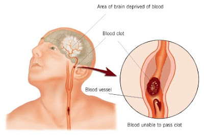
During a BBQ, a friend stumbled and took a little fall - she assured everyone that she was fine (they offered to call paramedics) and just tripped over a brick because of her new shoes. They got her cleaned up and got her a new plate of food - while she appeared a bit shaken up, Ingrid went about enjoying herself the rest of the evening. Ingrid's husband called later telling everyone that his wife had been taken to the hospital - (at 6:00pm, Ingrid passed away.) She had suffered a stroke at the BBQ. Had they known how to identify the signs of a stroke, perhaps Ingrid would be with us today. Some don't die. They end up in a helpless, hopeless condition instead.

It only takes a minute to read this...

A neurologist says that if he can get to a stroke victim within 3 hours he can totally reverse the effects of a stroke...totally. He said the trick was getting a stroke recognized, diagnosed, and then getting the patient medically cared for within 3 hours, which is tough.

RECOGNIZING A STROKE

Thank God for the sense to remember the "3" steps, STR . Read and Learn!

Sometimes symptoms of a stroke are difficult to identify. Unfortunately, the lack of awareness spells disaster. The stroke victim may suffer severe brain damage when people nearby fail to recognize the symptoms of a stroke.

Now doctors say a bystander can recognize a stroke by asking three simple questions:

S *Ask the individual to SMILE.

T *Ask the person to TALK . to SPEAK A SIMPLE SENTENCE
(Coherently) (ie. . It is sunny out today)

R *Ask him or her to RAISE BOTH ARMS.

{NOTE: Another 'sign' of a stroke is this: Ask the person to 'stick' out their tongue... if the tongue is 'crooked', if it goes to one side or the other that is also an indication of a stroke}

Tuesday, January 1, 2008

Appendicitis


Appendix
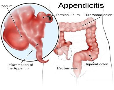
The appendix is a closed-ended, narrow tube up to several inches in length that attaches to the cecum (the first part of the colon) like a worm. (The anatomical name for the appendix, vermiform appendix, means worm-like appendage.) The inner lining of the appendix produces a small amount of mucus that flows through the open center of the appendix and into the cecum. The wall of the appendix contains lymphatic tissue that is part of the immune system for making antibodies. Like the rest of the colon, the wall of the appendix also contains a layer of muscle, but the muscle is poorly developed.

Appendicitis Appendicitis means inflammation of the appendix. It is thought that appendicitis begins when the opening from the appendix into the cecum becomes blocked. The blockage may be due to a build-up of thick mucus within the appendix or to stool that enters the appendix from the cecum. The mucus or stool hardens, becomes rock-like, and blocks the opening. This rock is called a fecalith (literally, a rock of stool). At other times, the lymphatic tissue in the appendix may swell and block the appendix. After the blockage occurs, bacteria which normally are found within the appendix begin to invade (infect) the wall of the appendix. The body responds to the invasion by mounting an attack on the bacteria, an attack called inflammation. An alternative theory for the cause of appendicitis is an initial rupture of the appendix followed by spread of bacteria outside the appendix.. The cause of such a rupture is unclear, but it may relate to changes that occur in the lymphatic tissue, for example, inflammation, that line the wall of the appendix.)

If the inflammation and infection spread through the wall of the appendix, the appendix can rupture. After rupture, infection can spread throughout the abdomen; however, it usually is confined to a small area surrounding the appendix (forming a peri-appendiceal abscess).

Sometimes, the body is successful in containing ("healing") the appendicitis without surgical treatment if the infection and accompanying inflammation do not spread throughout the abdomen. The inflammation, pain and symptoms may disappear. This is particularly true in elderly patients and when antibiotics are used. The patients then may come to the doctor long after the episode of appendicitis with a lump or a mass in the right lower abdomen that is due to the scarring that occurs during healing. This lump might raise the suspicion of cancer.

Symptoms
The main symptom of appendicitis is abdominal pain. The pain is at first diffuse and poorly localized, that is, not confined to one spot. (Poorly localized pain is typical whenever a problem is confined to the small intestine or colon, including the appendix.) The pain is so difficult to pinpoint that when asked to point to the area of the pain, most people indicate the location of the pain with a circular motion of their hand around the central part of their abdomen. A second, common, early symptom of appendicitis is loss of appetite which may progress to nausea and even vomiting. Nausea and vomiting also may occur later due to intestinal obstruction.

As appendiceal inflammation increases, it extends through the appendix to its outer covering and then to the lining of the abdomen, a thin membrane called the peritoneum. Once the peritoneum becomes inflamed, the pain changes and then can be localized clearly to one small area. Generally, this area is between the front of the right hip bone and the belly button. The exact point is named after Dr. Charles McBurney--McBurney's point. If the appendix ruptures and infection spreads throughout the abdomen, the pain becomes diffuse again as the entire lining of the abdomen becomes inflamed.

Diagnosis
The diagnosis of appendicitis begins with a thorough history and physical examination. Patients often have an elevated temperature, and there usually will be moderate to severe tenderness in the right lower abdomen when the doctor pushes there. If inflammation has spread to the peritoneum, there is frequently rebound tenderness. Rebound tenderness is pain that is worse when the doctor quickly releases his hand after gently pressing on the abdomen over the area of tenderness.

White Blood Cell Count
The white blood cell count in the blood usually becomes elevated with infection. In early appendicitis, before infection sets in, it can be normal, but most often there is at least a mild elevation even early. Unfortunately, appendicitis is not the only condition that causes elevated white blood cell counts. Almost any infection or inflammation can cause this count to be abnormally high. Therefore, an elevated white blood cell count alone cannot be used as a sign of appendicitis.

Urinalysis
Urinalysis is a microscopic examination of the urine that detects red blood cells, white blood cells and bacteria in the urine. Urinalysis usually is abnormal when there is inflammation or stones in the kidneys or bladder. The urinalysis also may be abnormal with appendicitis because the appendix lies near the ureter and bladder. If the inflammation of appendicitis is great enough, it can spread to the ureter and bladder leading to an abnormal urinalysis. Most patients with appendicitis, however, have a normal urinalysis. Therefore, a normal urinalysis suggests appendicitis more than a urinary tract problem.

Abdominal X-Ray
An abdominal x-ray may detect the fecalith (the hardened and calcified, pea-sized piece of stool that blocks the appendiceal opening) that may be the cause of appendicitis. This is especially true in children.

Ultrasound
An ultrasound is a painless procedure that uses sound waves to identify organs within the body. Ultrasound can identify an enlarged appendix or an abscess. Nevertheless, during appendicitis, the appendix can be seen in only 50% of patients. Therefore, not seeing the appendix during an ultrasound does not exclude appendicitis. Ultrasound also is helpful in women because it can exclude the presence of conditions involving the ovaries, fallopian tubes and uterus that can mimic appendicitis.

Barium Enema
A barium enema is an x-ray test where liquid barium is inserted into the colon from the anus to fill the colon. This test can, at times, show an impression on the colon in the area of the appendix where the inflammation from the adjacent inflammation impinges on the colon. Barium enema also can exclude other intestinal problems that mimic appendicitis, for example Crohn's disease.

Computerized tomography (CT) Scan
In patients who are not pregnant, a CT Scan of the area of the appendix is useful in diagnosing appendicitis and peri-appendiceal abscesses as well as in excluding other diseases inside the abdomen and pelvis that can mimic appendicitis.

Laparoscopy
Laparoscopy is a surgical procedure in which a small fiberoptic tube with a camera is inserted into the abdomen through a small puncture made on the abdominal wall. Laparoscopy allows a direct view of the appendix as well as other abdominal and pelvic organs. If appendicitis is found, the inflamed appendix can be removed with the laparascope. The disadvantage of laparoscopy compared to ultrasound and CT is that it requires a general anesthetic.

There is no one test that will diagnose appendicitis with certainty. Therefore, the approach to suspected appendicitis may include a period of observation, tests as previously discussed, or surgery.

Treatment
Once a diagnosis of appendicitis is made, an appendectomy usually is performed. Antibiotics almost always are begun prior to surgery and as soon as appendicitis is suspected.

There is a small group of patients in whom the inflammation and infection of appendicitis remain mild and localized to a small area. The body is able not only to contain the inflammation and infection but to resolve it as well. These patients usually are not very ill and improve during several days of observation. This type of appendicitis is referred to as "confined appendicitis" and may be treated with antibiotics alone. The appendix may or may not be removed at a later time.

On occasion, a person may not see their doctor until appendicitis with rupture has been present for many days or even weeks. In this situation, an abscess usually has formed, and the appendiceal perforation may have closed over. If the abscess is small, it initially can be treated with antibiotics; however, the abscess usually requires drainage. A drain (a small plastic or rubber tube) usually is inserted through the skin and into the abscess with the aid of an ultrasound or CT scan that can determine the exact location of the abscess. The drain allows pus to flow from the abscess out of the body. The appendix may be removed several weeks or months after the abscess has resolved. This is called an interval appendectomy and is done to prevent a second attack of appendicitis.

Surgical Intervention: Appendectomy
During an appendectomy, an incision two to three inches in length is made through the skin and the layers of the abdominal wall over the area of the appendix. The surgeon enters the abdomen and looks for the appendix which usually is in the right lower abdomen. After examining the area around the appendix to be certain that no additional problem is present, the appendix is removed. This is done by freeing the appendix from its mesenteric attachment to the abdomen and colon, cutting the appendix from the colon, and sewing over the hole in the colon. If an abscess is present, the pus can be drained with drains that pass from the abscess and out through the skin. The abdominal incision then is closed.

Newer techniques for removing the appendix involve the use of the laparoscope. The laparoscope is a thin telescope attached to a video camera that allows the surgeon to inspect the inside of the abdomen through a small puncture wound (instead of a larger incision). If appendicitis is found, the appendix can be removed with special instruments that can be passed into the abdomen, just like the laparoscope, through small puncture wounds. The benefits of the laparoscopic technique include less post-operative pain (since much of the post-surgery pain comes from incisions) and a speedier return to normal activities. An additional advantage of laparoscopy is that it allows the surgeon to look inside the abdomen to make a clear diagnosis in cases in which the diagnosis of appendicitis is in doubt. For example, laparoscopy is especially helpful in menstruating women in whom a rupture of an ovarian cysts may mimic appendicitis.

If the appendix is not ruptured (perforated) at the time of surgery, the patient generally is sent home from the hospital after surgery in one or two days. Patients whose appendix has perforated are sicker than patients without perforation, and their hospital stay often is prolonged (four to seven days), particularly if peritonitis has occurred. Intravenous antibiotics are given in the hospital to fight infection and assist in resolving any abscess.

Occasionally, the surgeon may find a normal-appearing appendix and no other cause for the patient's problem. In this situation, the surgeon may remove the appendix. The reasoning in these cases is that it is better to remove a normal-appearing appendix than to miss and not treat appropriately an early or mild case of appendicitis.