Sunday, May 20, 2007
Ayurveda आयुर्वेद
Ayurveda (Devanagari: आयुर्वेद ) or Ayurvedic medicine is an ancient system of health care that is native to the Indian subcontinent. The word "Ayurveda" is a tatpurusha compound of the word āyus meaning "life" or "life principle", and the word veda, which refers to a system of "knowledge". Thus "Ayurveda" roughly translates as the "knowledge of life". According to Charaka, "life" itself is defined as the "combination of the body, sense organs, mind and soul, the factor responsible for preventing decay and death, which sustains the body over time, and guides the processes of rebirth" [1]. According to this perspective, Ayurveda is concerned with measures to protect "ayus", which includes healthy living along with therapeutic measures that relate to physical, mental, social and spiritual harmony. Ayurveda is also one among the few traditional systems of medicine to contain a sophisticated system of surgery (which is referred to as "salya-chikitsa").
According to the Ayurvedavatarana (the "descent of Ayurveda"), the origin of Ayurveda is stated to be a divine revelation of the ancient Indian creator Hindu God Lord Brahma.[2] as he awoke to recreate the universe. This knowledge was passed directly to Daksha Prajapati in the form of shloka sung by Lord Brahma.[3], and this was in turn passed down through a successive chain of deities to God Indra, the protector of dharma. According to this account, the first human exponent of Ayurveda was Bharadvaja, who learned it directly from Indra. Bharadvaja in turn taught Ayurveda to a group of assembled sages, who then passed down different aspects of this knowledge to their students. According to tradition, Ayurveda was first described in text form by Agnivesha, in his book the Agnivesh tantra. The book was later redacted by Charaka, and became known as the Charaka Samhitā.[4] Another early text of Ayurveda is the Sushruta Samhitā, which was compiled by Sushrut, the primary pupil of Dhanvantri, sometime around 1000 B.C.E.. Dhanvantri is known as the Father of Surgery, and in the Sushrut Samhita, the teachings and surgical techniques of Dhanvantri are compiled and complemented with additional findings and observations of Sushrut regarding topics ranging from obstetrics and orthopedics to ophthalmology. Sushrut Samhita together with Charaka Samhitā, served as the textual material within the ancient Universities of Takshashila and Nalanda.[5] These texts are believed to have been written around the beginning of the Common Era, and is based on a holistic approach rooted in the philosophy of the Vedas and Vedic culture.
Alzheimer Disease (AD)
Frequency:
* In the US: The lifetime risk of AD is estimated to be 1:4-1:2. More than 14% of individuals older than 65 years have AD, and the prevalence increases to at least 40% in individuals older than 80 years.
* Internationally: Prevalences similar to those in the United States have been reported in industrialized nations. Countries experiencing rapid increases in the elderly segments of their population have rates approaching those in the United States.
Mortality/Morbidity:
* Second to only certain cancers and cardiovascular disease, AD is frequently considered a leading cause of death in the United States.
* The primary cause of death is intercurrent illness, such as pneumonia, in a patient who has experienced the debilitating effects of AD for many years.
Race: Some claim that AD affects certain ethnic and racial groups more severely than others, but more study is needed before reliable statements about racial predilections can be made.
Sex: AD affects both men and women. Many studies indicate that the risk of AD is significantly higher in women than in men. Some authorities have postulated that this difference is due to the loss of the neurotrophic effect of estrogen in postmenopausal women. Other factors may also influence this relative difference.
Age: The prevalence of AD increases with age.
* AD is most prevalent in individuals older than 60 years. Some forms of familial early-onset AD can appear as early as the third decade, but this represents a subgroup of the less than 10% of all familial cases of AD.
* More than 90% of cases of AD are sporadic and occur in individuals older than 60 years.
* Of interest, results of some studies of nonagenarians and centenarians suggest that the risk decreases in individuals older than 80 or 90 years. If so, age is not an unqualified risk factor for the disease, but further study of this matter is needed.
Friday, May 18, 2007
Back Pain
of low back pain, with more than half of that spent on surgical treatment.
The lower back is a well designed construction of bone, muscle and ligament. Your backbone (vertebral column) is actually a stack of more than 30 small bones called vertebrae. Together, they create a bony canal that surrounds and protects your spinal cord. Small nerves enter and exit the spinal canal through spaces in your vertebrae called the foramen.
These vertebrae are held together by muscles, tendons and ligaments. Between the vertebrae are discs, which act as "shock absorbers" and prevent the vertebrae from hitting one another when you walk, run or jump. They also allow your spine to twist, bend and extend. Since the lower back is the hinge between the upper and lower body and carries most of your weight, it is especially vulnerable to injury and is the site of most back pain. When low back pain strikes, we become acutely aware of just how much we rely on a flexible, strong back.
The Casues of Back Pain
The most common causes of low back pain are:
Pinched Nerves - Pressure or impingement on nerve roots in the spinal canal can be caused by:
Herniated Disc - A herniated disc, (also called bulging disc or slipped disc) is a rupture often brought on by repeated vibration or motion (as during machine use or sport activity, or when lifting improperly), or by a sudden heavy strain or increased pressure to the lower back. Back pain and leg pain can result when the herniated disc pinches one of the nerves. A herniated disc in the lumbar region can affect the nerves, which runs from your spinal cord to your leg. Compression or inflammation of this nerve causes sciatica - a sharp, shooting pain in your lower back, buttocks and leg.
Degenerative Disc Disease - As we age, the water and protein content of the body's cartilage changes. This change results in weaker, more fragile and thin cartilage. Because both the discs and the joints that stack the vertebrae (facet joints) are partly composed of cartilage, these areas are subject to wear and tear over time (degenerative changes). The gradual deterioration of the disc between the vertebrae is referred to as Degenerative Disc Disease.
Bone Spurs - also termed osteophytes (os-tee-o-fights). Osteophytes may be found in areas affected by arthritis such as the disc or joint spaces where cartilage has deteriorated. The body's production of osteophytes is a futile attempt to stop the motion of the arthritic joint and deal with the degenerative process. It never completely works. The evidence of bony deposits can be found on an x-ray or MRI. A bone spur may cause nerve impingement at the neuroforamen (nu-row for-a-men). The neuroforamen are passageways through which the nerve roots exit the spinal canal. Sensory symptoms include pain, numbness, burning and pins and needles in the extremities below the affected spinal nerve root. Motor symptoms include muscle spasm, cramping, weakness, or loss of muscular control in a part of the body.
Spinal Stenosis is the narrowing of the spinal canal by a piece of bone, ligamentus flavus thickening or disc material. This can cause weakness in your extremities and typically develops with age.
Osteoporosis, which means "porous bones," causes bones to become weak and brittle - so brittle that even mild stresses like bending over, lifting a vacuum cleaner or coughing can cause a fracture. In most cases, bones weaken when you have low levels of calcium, phosphorus and other minerals in your bones. The pain from an osteoporotic spinal fracture can last for several weeks as the bone heals, and then typically turns into more of a chronic, achy pain concentrated in the area of the back where the fracture occurred. This aching may be similar to the sensation reported by those with osteoarthritis. A bone density test, which measures bone mass, preferably taken of both a long bone and a vertebral body, is used to diagnose osteoporosis.
Osteoarthritis or Facet Disease is a degenerative joint condition that causes slow deterioration of cartilage. Osteoarthritis of the spine results in narrowed cartilage disks between the bones that make up your backbone. Without this cartilage cushioning, the joints (facets) between adjacent bones compress and become irregular, causing inflammation, pain, swelling and stiffness. Your body tries to compensate for this form of arthritis, but the repairs are often inadequate, resulting in little growths of additional bone called bone spurs.
Cervical spondylosis is a common condition that results from degeneration (osteoarthritis) of the bones of the neck (cervical vertebrae). This can lead to increasing pain in the neck and arm, weakness, and changes in sensation.
Spinal deformities such as scoliosis, which is an abnormal curvature of the spine. Most cases are mild, but severe cases may require treatment with braces or surgery.
Small injury to a muscle (strain) or a ligament (sprain) from improper lifting, excess body weight and poor posture. Strains and sprains can also develop from carrying heavy handbags or briefcases or sleeping in an awkward position.
Compression fractures, more common among postmenopausal women with osteoporosis, or after long-term corticosteroid use. In a person with osteoporosis, even a small amount of force put on the spine, as from a sneeze, may cause a compression fracture.
Fractures of the vertebrae caused by significant force (as from an auto or bicycle accident, a direct blow to the spine, or compressing the spine by falling onto the buttocks or head).
Most pain in the low back (lumbar region) is triggered by some combination of overuse, muscle strain, or injury to the muscles and ligaments that support the spine. Many experts believe that over time, chronic muscle strain can lead to an overall imbalance in the spinal structure. This leads to a constant tension on the muscles, ligaments, bones, and discs, making the back more prone to injury or re-injury.
The causes of low back pain tend to be interrelated. For example, after straining muscles, you are likely to use your back differently than usual. As other parts of your back work harder or move in unaccustomed ways to make up for the injured muscles, they also become more prone to injury.
The Symptoms of Back Pain
Back pain can be:
Acute, lasting less than 3 months. Most people gain relief after 4 to 6 weeks of home treatment.
Recurrent, a repeat episode of acute symptoms. Most people have at least one episode of recurrent low back pain.
Chronic, lasting longer than 3 months.
The term "low back pain" is used to describe a spectrum of symptoms. Depending on the cause, low back pain may be dull, burning, or sharp, covering a broad area or confined to a single point. It can come on gradually or suddenly and may or may not be accompanied by muscle spasms or stiffness.
Leg symptoms can be caused by lower spine problems that place pressure on a nerve to the leg; they can occur on their own or along with low back pain. Leg symptoms can include pain, numbness, or tingling, usually below the knee.
Weakness in both legs, along with loss of bladder and/or bowel control, are symptoms of cauda equine syndrome, which requires immediate medical attention.
Numbness and tingling are felt when nerve impulses aren't traveling properly from the skin to the brain. A patient with back problems may also experience numbness in other parts of the body, especially the legs and feet. This always indicates some kind of nerve damage in the peripheral nervous system or the central nervous system (i.e. the spine or the brain) and requires prompt and serious attention.
Common causes of numbness include the following:
Radiculopathy - A pinched nerve due to a herniated disc.
Stenosis - A narrowing of the spinal canal, which can compress sensory nerve fibers causing loss of sensation.
Multiple Sclerosis
Stroke, and
Diabetes
Treatment of Back Pain
Your health professional can assess acute low back pain by talking to you about your medical history and your work and physical activities, and doing a simple physical examination. For 95% of people with low back pain, this type of assessment is all that is necessary for a health professional to make treatment recommendations
Monday, May 14, 2007
Over weight Problem? Here is an Answer
Think closely about what we're about to tell you, since it's going to change the way you think about dieting...
FOOD is more powerful than any prescription weight loss pills, because the FOOD that you eat can either make you THIN or FAT. You don't get fat because of a lack of exercising, that's a myth. You get fat because you don't eat the right foods at the right intervals each day.
Also, the pattern that you choose to eat your meals each day is more powerful than any prescription weight loss pills. This is true because your body is like an "engine" and it only needs certain foods at certain intervals each day, and if you don't eat the right foods at the right times then it won't burn those calories -- and you'll wind up storing those calories as fat tissue. (Hint: You need to eat more than 3 times per day to lose weight, but we'll show you the details later).
You have gotten overweight by eating the wrong foods, that much is a fact. And guess what? You can get SLIM by eating the RIGHT FOODS at the RIGHT INTERVALS each day.
It's not really any more complicated than that, and the way to start losing weight has nothing to do with starving yourself or jogging.
The reason you cannot lose weight by starving yourself (using a low calorie diet) is because your metabolism will detect any major drop in calories and it will then ADJUST ITSELF by burning fewer calories each day.
For example:
If you begin eating 2,500 calories per day then your metabolism will adjust itself so that your body begins burning 2,500 calories per day.
If you try to starve yourself by suddenly eating 1,000 calories per day then your metabolism will again ADJUST ITSELF so that your body begins to burn only 1,000 calories per day. That's why you have failed in your past dieting attempts, that's why you always seem to fail when you try and starve yourself.
Now you know the reason why you can eat 1,000 calories per day and not lose any weight while your friends can eat 2,500 calories per day and not gain any weight.
Also, virtually every person in today's society is buying mostly "low fat" or "non fat" food at the grocery store, everybody is conscious of the "fat grams" inside the food they buy. However, people are getting fatter than ever by doing this and people are not losing weight by switching to the "low fat lifestyle".
Finally, the ridiculous "low carb" diets have become popular in recent years, but of course those are just thinly disguised starvation diets. If you don't think so then check out these facts below about Atkins....
The Atkins Diet limits you to ONLY 20 grams of "net carbs" per day during the first phase of the diet......which means that you cannot even eat a single large apple during the first phase of the Atkins Diet (since even a large apple has more than 20 grams of net carbs).
...And if you want to continue through all of the phases of the Atkins Diet, then it will take another 4 MONTHS (16 weeks) before you'll be allowed to eat just 400 carb calories per day -- and any typical restaurant meal has more carbs than that. So this is really just another form of starving and torturing yourself, which is not the answer.
The Shifting Calories Theory...
Your metabolism doesn't know how much food you'll eat tomorrow or the next day because those days have not happened yet.
Therefore, your metabolism always burns calories based on your eating habits during the past few days -- because it assumes that you'll continue to eat in the same general way.
Guess what? You're about to shock your metabolism by doing something you've never tried before -- you're going to do the OPPOSITE of what it expects you to do. You're going to NOT continue eating the same types of calories and meals for more than a couple days at a time, and you're going to lose a lot of weight by doing this.
To make this work you need to SHIFT the types of calories eaten as shown in our diet on the next page, and if you do this then your metabolism will burn all of the calories eaten. Then, when it finishes burning those calories it will find the nearest available fat tissue on your body and burn that too...
To lose weight your diet menu needs to be SHIFTED every few days -- and this is something you've never tried before, and that's why you've never been able to change your body when dieting.
Of course, in order for this dieting technique to work you also need to eat foods which have been rated "Fat Burning Compatible" -- because the foods you eat must be easy to burn (so that your metabolism will burn them entirely and then switch to burning your fat tissue after that).
Sunday, May 6, 2007
Blood Pressure (BP) Test
Test Overview
Blood pressure is a measure of the force of blood inside an artery. A blood pressure measurement is taken by temporarily stopping the flow of blood in an artery (usually by inflating a cuff around the upper arm) and then listening for the sound of the blood beginning to flow through the artery again as air is released from the cuff.
As blood flows through the artery, it can be heard through a stethoscope placed on the skin over the artery. Blood pressure is recorded as two measurements.
- The reading on the gauge when blood flow is first heard is called the systolic pressure. Systolic pressure represents the peak blood pressure that occurs when the heart contracts.
- The reading on the gauge when blood flow can no longer be heard is the diastolic pressure. Diastolic pressure represents the lowest blood pressure that occurs when the heart relaxes between beats.
These two pressures are expressed in millimeters of mercury (mm Hg) because the original devices that measured blood pressure used a column of mercury. Blood pressure measurements are recorded as systolic/diastolic. For example, if your systolic pressure is 120 mm Hg and your diastolic pressure is 80 mm Hg, your blood pressure is recorded as 120/80 and read as "120 over 80."
Level | ||
High blood pressure is: | 140 or above | 90 or above |
Prehypertension is: | 120 to 139 | 80 to 89 |
Normal adult (age 18 or older) blood pressure is: | 119 or below | 79 or below |
High blood pressure means that the heart must work harder to pump blood throughout the body. Uncontrolled high blood pressure increases the risk of stroke, heart failure, kidney failure, and heart attack. As blood pressure increases, the risk of it causing these problems also increases.
High blood pressure is often referred to as the "silent killer," because it usually has no obvious symptoms and most people cannot tell whether their own blood pressure is high unless it is measured.
High blood pressure in pregnancy
Your health professional will monitor your blood pressure during pregnancy by measuring it at every prenatal visit, and you can also monitor it at home. Rapidly increasing or abnormally high blood pressure can be a sign of a pregnancy-related condition that affects the mother's kidneys, liver, and brain, and the placenta (preeclampsia). Because an increase in blood pressure can be a first sign of preeclampsia, you will be closely monitored if you have elevated blood pressure. Severe high blood pressure can be dangerous to both you and your baby. If you have chronic high blood pressure, your blood pressure will be closely monitored during your entire pregnancy.
Home blood pressure monitors
Home blood pressure monitors make it easy to measure your blood pressure at home. If you are concerned that you might have high blood pressure or if your family has a history of high blood
pressure, you may want to consider getting a home blood pressure monitor. If you know you have high blood pressure, you may want to get one to monitor your response to any blood pressure medication you are taking regularly.
The two general types of blood pressure monitors commonly available are manual and automatic. (Automatic types may also be called electronic or digital.)
Manual blood pressure monitors
Manual models are similar to those that your health professional might use to take your blood pressure. Called a sphygmomanometer, these devices usually include an arm cuff, a squeeze bulb for inflation, a stethoscope or microphone, and a mechanical gauge to measure the blood pressure. Manual blood pressure monitors require good eyesight and hearing to use them correctly. There are two basic styles of manual blood pressure devices.
These display the blood pressure on a circular dial with a needle. As the pressure in the cuff rises, the needle moves clockwise on the dial. As the cuff pressure falls, the needle moves counterclockwise. Again, a stethoscope is required; some models have the stethoscope head permanently attached to the cuff. The aneroid devices are compact and inexpensive but somewhat difficult to use. Also, the dial gauges may need to be recalibrated from time to time to maintain their accuracy.
Automatic (also called electronic or digital) blood pressure monitors
Electronic battery-operated monitors use a microphone to detect blood pulsing in the artery instead of having to listen with a stethoscope. The cuff, which is attached to your wrist or upper arm, is connected to an electronic monitor that automatically inflates and deflates the cuff when you press the start button. First you place your wrist or upper arm inside the cuff. Then press the start button on the monitor and wait for a reading to display. The monitor records your pulse as well as your blood pressure.
The electronic devices are by far the easiest to use, but they are also the most expensive. Generally, the electronic models that use an arm cuff are more accurate than those that use a wrist cuff.
The type of blood pressure monitor typically found in supermarkets, pharmacies, and shopping malls is an electronic device.
Ambulatory blood pressure monitoring (ABPM)
Another method of measuring blood pressure, called ambulatory blood pressure monitoring (ABPM), may be ordered by your health professional to provide a more accurate picture of your blood pressure over time. ABPM may be done if a manual or an electronic method of measurement yields inconsistent results.
ABPM automatically records blood pressure over a period of a few hours to an entire day. The device generally consists of a cuff worn on one arm and a monitor worn around the waist. Your health professional's office will fit you with the monitor and provide instructions on its use.
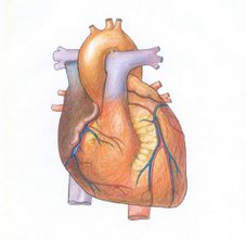
Cardiac Arrest/ Heart Attack
A cardiac arrest, or circulatory arrest, is the abrupt cessation of normal circulation of the blood due to failure of the heart to contract effectively during systole.[1]
"Arrested" blood circulation prevents delivery of oxygen to all parts of the body. Cerebral hypoxia, or lack of oxygen supply to the brain, causes victims to lose consciousness and to stop normal breathing. Brain injury is likely if cardiac arrest is untreated for more than 5 minutes,[2] although new treatments such as induced hypothermia have begun to extend this time.[3][4] To improve survival and neurological recovery immediate response is paramount.[5]
Cardiac arrest is a medical emergency that, in certain groups of patients, is potentially reversible if treated early enough (See Reversible Causes, below). When unexpected cardiac arrest leads to death this is called sudden cardiac death (SCD).[1] The primary first-aid treatment for cardiac arrest is cardiopulmonary resuscitation (commonly known as CPR) to provide circulatory support until availability of definitive medical treatment, which will vary dependant on the rhythm the heart is exhibiting, but often requires Defibrillation.
Causes of Cardiac Arrest
Ventricular fibrillation (VF) constitutes the most common electrical mechanism in cardiac arrest, and is responsible for 65 to 80% of occurrences. Another 20-30% is caused by severe bradyarrhythmias, pulseless electrical activity (PEA) and asystole. Other conditions are associated with impaired circulation due to a state of shock.[1]
Among adults ischemic heart disease is the predominant cause.[6] At autopsy 30% of victims show signs of recent myocardial infarction. Other conditions include structural abnormalities, arrhythmias and cardiomyopathies. Secondary cardiac arrest may be elicited by non-cardiac conditions such as hypoxia from a variety of causes,[7] overwhelming infection (sepsis), pulmonary embolism, arrythmias, cardiac tamponade, shock, pneumothorax, ventricular rupture, as well as other conditions such as electrocution and near-drowning. Non-cardiac conditions constitute the principal cause of cardiac arrest in in-hospital patients.[8]
Coronary heart disease (CHD) -also known as coronary artery disease, or (CAD)- is the predominant disease process associated with sudden cardiac death in the United States and elsewhere in the developed world. The incidence of CHD in individuals who suffer sudden cardiac death is between 64 and 90%.
In children, cardiac arrest is typically caused by hypoxia from other causes such as near-drowning. With prompt treatment survival rates are high.
Treatable causes
The most important treatable cause of cardiac arrest is ventricular fibrillation. The only definitive treatment is defibrillation, although the entire chain of survival must be intact if a victim of out-of-hospital cardiopulmonary arrest is to survive. The chain of survival consists of 1. early access to emergency medical services, 2. early CPR, 3. early defibrillation, and 4. early advanced care. The efficacy of defibrillation is time dependent, and the odds of successful resuscitation decline rapidly from the onset of cardiopulmonary arrest. However, current research suggests that 1.5 to 3 minutes of CPR prior to the first shock may increase survival rates when down times exceed 4 minutes and no CPR has been performed prior to the defibrillator's arrival.
For other causes of cardiac arrest, the best treatment is prevention. Aggressive therapy innitiated in the peri-arrest period may prevent cardiac arrest and subsequent death. Reversible causes of asystole and pulseless electrical activity include the "Hs and Ts.